DRI OCT Triton, Swept Source OCT
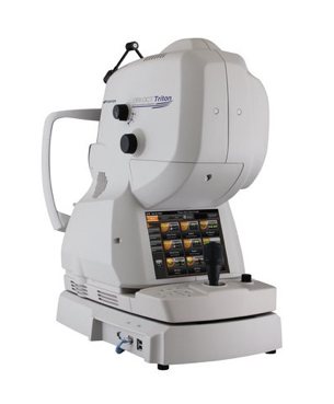
DRI OCT Triton, prednji i zadnji Swept Source OCT sa ugrađenom funkcijom snimanja fundusa pomoću nemidrijatične fundus kamere
Ključne karakteristike
- Pouzdanija početna dijagnoza i sposobnost praćenja promena tokom operacije
- Bolja klinička efikasnost i praćenje pacijenta
- Bolja udobnost pacijenata
- Jednokratno skeniranje i pohrana celokupne informacije
Originalni tekst i dodatno pojašnjenje preuzeli smo sa Topcon Europe Medical internet stranice!
Topcon is the first in the world to introduce a combined anterior & posterior Swept Source OCT, the DRI OCT Triton. The DRI OCT Triton incorporates full color high resolution fundus photography and FA & FAF imaging. FA & FAF imaging is a factory option.
Swept Source technology & 1,050nm wave length
Swept Source OCT provides a significant improvement over conventional OCT. Due to the optimized long wavelength scanning light (1,050nm), there is better penetration of the deeper layers of the eye. Furthermore, this scanning light also penetrates better through cataracts, hemorrhages, blood vessels and sclera.
The world’s fastest scanning speed 100,000 A-Scans/second
Approximately twice higher scan speed, compared to Topcon SD OCT, will bring more scans for a single B-scan image, and more informative image supports efficiency and quality of diagnosis.
Better penetration
The high penetration of the Swept Source light can easily and clearly visualize deep layers in the eye, such as choroid and sclera. A further benefit of Swept Source is that it can clearly visualize both the vitreous and choroid in a single scan, that are uniformly clear and noise-free. This eliminates the need for time consuming vitreous/choroidal combination scans.
Wide and deep scans
In one single image the vitreous & choroid are revealed in a crystal clear way. The Topcon DRI OCT Triton enhances visualization of outer retinal structures and deep pathologies. The Topcon DRI OCT Triton automatically detects 7 boundaries including the chorio-scleral interface. The 12mm B-scan covers both the macular area and the optic disc.
Invisible scan lines
The invisible 1,050nm wavelength does not distract patients. Patients do not see the scanning line, which is an advantage with elderly patients and children. Reduction in movement artifacts and increased repeatability.
Time efficiency - create one single overview
Combination scans cover the macular and disc areas in a single shot, and offer both macular and Retinal Nerve Fiber Layer (RFNL) analysis. Combination scans are time efficient for the operator and convenient for the patient. Combination scans allow both macular and disc analysis in one overview.
Multi modal fundus imaging
The Topcon DRI OCT Triton offers a true color, non mydriatic fundus image while using a very low intensity flash. This unique feature is a perfect tool for identifying the location of scans in the eye utilizing TOPCON’s patented Pinpoint RegistrationTM. The DRI OCT Triton Plus offers a wide range of diagnostic options with multi-modal color fundus imaging, Fluorescein Angiography (FA) and Fundus Autofluorescence (FAF) for even more diagnostic possibilities. For the first time Pinpoint registrationTM will be available with fundus auto fluorescence and Swept Source OCT.
New tracking system - SMARTTrackTM
SMARTTrackTM is a very useful tool to compensate for the ever present involuntary eye movements (microsaccades). It allows the automatic acquisition of a follow-up scan in precisely the same anatomical location. SMARTTrackTM enhances the user-friendliness of the machine.
Anterior segment analysis
The Topcon DRI OCT Triton can be extended to include anterior imaging, making the Swept Source a versatile diagnosis tool for both anterior and posterior imaging. The anterior attachment ensures sharp images, even in the periphery of the cornea and in depth images of the anterior chamber.
